Upper Leg Tendon Anatomy : Muscles Of The Anterior Thigh Quadriceps Teachmeanatomy : Upper limb trauma programme of extensor tendons are essential in the rehabilitation of these types of injuries.
byAdmin-
0
Upper Leg Tendon Anatomy : Muscles Of The Anterior Thigh Quadriceps Teachmeanatomy : Upper limb trauma programme of extensor tendons are essential in the rehabilitation of these types of injuries.. Related posts of muscle anatomy upper leg. The patella is a large sesamoid (a bone within a tendon) bone the medial and lateral parts of quadriceps femoris descend on either side of the patella and are inserted onto the upper anterior surface of the tibia. Flexibility of the plantar flexors was related to nvo7 (+0.38, p = 0.05). The patellar tendon runs inferiorly from the patella bone to the tibial tuberosity. Human forearm anatomy inside arm anatomy upper arm anatomy skin left lower arm anatomy leg muscle and tendon anatomy arm anatomy names arm parts anatomy anterior arm muscle anatomy upper arm muscle tear lateral of upper arm muscle anatomy upper arm muscles.
There is no real division between the core and the upper leg; Concept conceptual 3d illustration fit strong back upper leg human anatomy, anatomical muscle isolated white background for body medical health tendon foot and biological gym fitness muscular system. Tendon, tissue that attaches a muscle to other body parts, usually bones. The human leg, in the general word sense, is the entire lower limb of the human body, including the foot, thigh and even the hip or gluteal region. Human forearm anatomy inside arm anatomy upper arm anatomy skin left lower arm anatomy leg muscle and tendon anatomy arm anatomy names arm parts anatomy anterior arm muscle anatomy upper arm muscle tear lateral of upper arm muscle anatomy upper arm muscles.
Tensor Fasciae Latae Muscle Wikipedia from upload.wikimedia.org Achilles tendon cross section was not related to walking or running economy. The posterior talofibular ligament is attached to the posterolateral tubercle, which is larger and more prominent than the posteromedial tubercle. N., morris s.f., hallock g.g., neligan p.c. Tendons transmit the mechanical force of muscle contraction to the bones. Collectively, the muscles in this area plantarflex and invert the foot. Upper limb trauma programme of extensor tendons are essential in the rehabilitation of these types of injuries. The pads of the machine are situated at the achilles tendon. In this upper leg tutorial, i go over all the major points of the upper leg to take your sculpting skills.
The sulcus for this tendon is flanked by the posterolateral and posteromedial tubercles.
The pads of the machine are situated at the achilles tendon. Human forearm anatomy inside arm anatomy upper arm anatomy skin left lower arm anatomy leg muscle and tendon anatomy arm anatomy names arm parts anatomy anterior arm muscle anatomy upper arm muscle tear lateral of upper arm muscle anatomy upper arm muscles. • transmit away from cell body. The tendons for these muscles begin at your ischial tuberosity, or ischium (the. An anatomical and biomechanical study. We study anatomy at the practical anatomy class we study the human body. It serves to attach the plantaris, gastrocnemius (calf) and soleus muscles to the calcaneus (heel) bone. The achilles tendon or heel cord, also known as the calcaneal tendon, is a tendon at the back of the lower leg, and is the thickest in the human body. Choose from 500 different sets of flashcards about anatomy muscle anatomy_ upper leg on quizlet. Muscle/tendon inflammation and pain along anterio… Tendon, tissue that attaches a muscle to other body parts, usually bones. The patellar tendon runs inferiorly from the patella bone to the tibial tuberosity. We speak of the upper extremities (arms) and the lower extremities (legs).
How does achilles tendon rupture occur… why are achilles piercings dangerous? The calf comprises of 2 major muscles (gastrocnemius and soleus) both of which insert into the heel bone via the achilles tendon. Tendon, tissue that attaches a muscle to other body parts, usually bones. Tendons transmit the mechanical force of muscle contraction to the bones. Flexibility of the plantar flexors was related to nvo7 (+0.38, p = 0.05).
Anatomy Leg Muscles Diagram Quizlet from o.quizlet.com Current techniques have tended to anatomical reconstruction of the lcl, pt and pf. How does achilles tendon rupture occur… why are achilles piercings dangerous? N., morris s.f., hallock g.g., neligan p.c. The tendons for these muscles begin at your ischial tuberosity, or ischium (the. Spicermanyt at checkout for 40% off this tutorial! Mnemonics that can be used to remember the anatomy of the ankle tendons from anterior to posterior as they pass posteriorly to the medial malleolus of the tibia under the flexor retinaculum in the tarsal tunnel include: Superficial veins of upper limb , anatomy : Tendons are also bands of connective tissue.
By spicer mcleroy in tutorials.
The posterior talofibular ligament is attached to the posterolateral tubercle, which is larger and more prominent than the posteromedial tubercle. Collectively, the muscles in this area plantarflex and invert the foot. Mnemonics that can be used to remember the anatomy of the ankle tendons from anterior to posterior as they pass posteriorly to the medial malleolus of the tibia under the flexor retinaculum in the tarsal tunnel include: Spicermanyt at checkout for 40% off this tutorial! The patella is a large sesamoid (a bone within a tendon) bone the medial and lateral parts of quadriceps femoris descend on either side of the patella and are inserted onto the upper anterior surface of the tibia. Hands are outstretched, holding onto the handles of the bench. The achilles tendon or heel cord, also known as the calcaneal tendon, is a tendon at the back of the lower leg, and is the thickest in the human body. There is no real division between the core and the upper leg; Concept conceptual 3d illustration fit strong back upper leg human anatomy, anatomical muscle isolated white background for body medical health tendon foot and biological gym fitness muscular system. 17.03.2021 · upper leg tendon anatomy : The patellar tendon runs inferiorly from the patella bone to the tibial tuberosity. Flexibility of the plantar flexors was related to nvo7 (+0.38, p = 0.05). Localized anatomy of the hamstring muscles including semimembranosus, semitendinosus, biceps the hamstrings refer to 3 long posterior leg muscles, the biceps femoris, semitendinosus, and semimembranosus.
The achilles tendon or heel cord, also known as the calcaneal tendon, is a tendon at the back of the lower leg, and is the thickest in the human body. ✓ quadriceps tendon attached superior and patellar ligament inferior. In this upper leg tutorial, i go over all the major points of the upper leg to take your sculpting skills. Related posts of muscle anatomy upper leg. The sulcus for this tendon is flanked by the posterolateral and posteromedial tubercles.
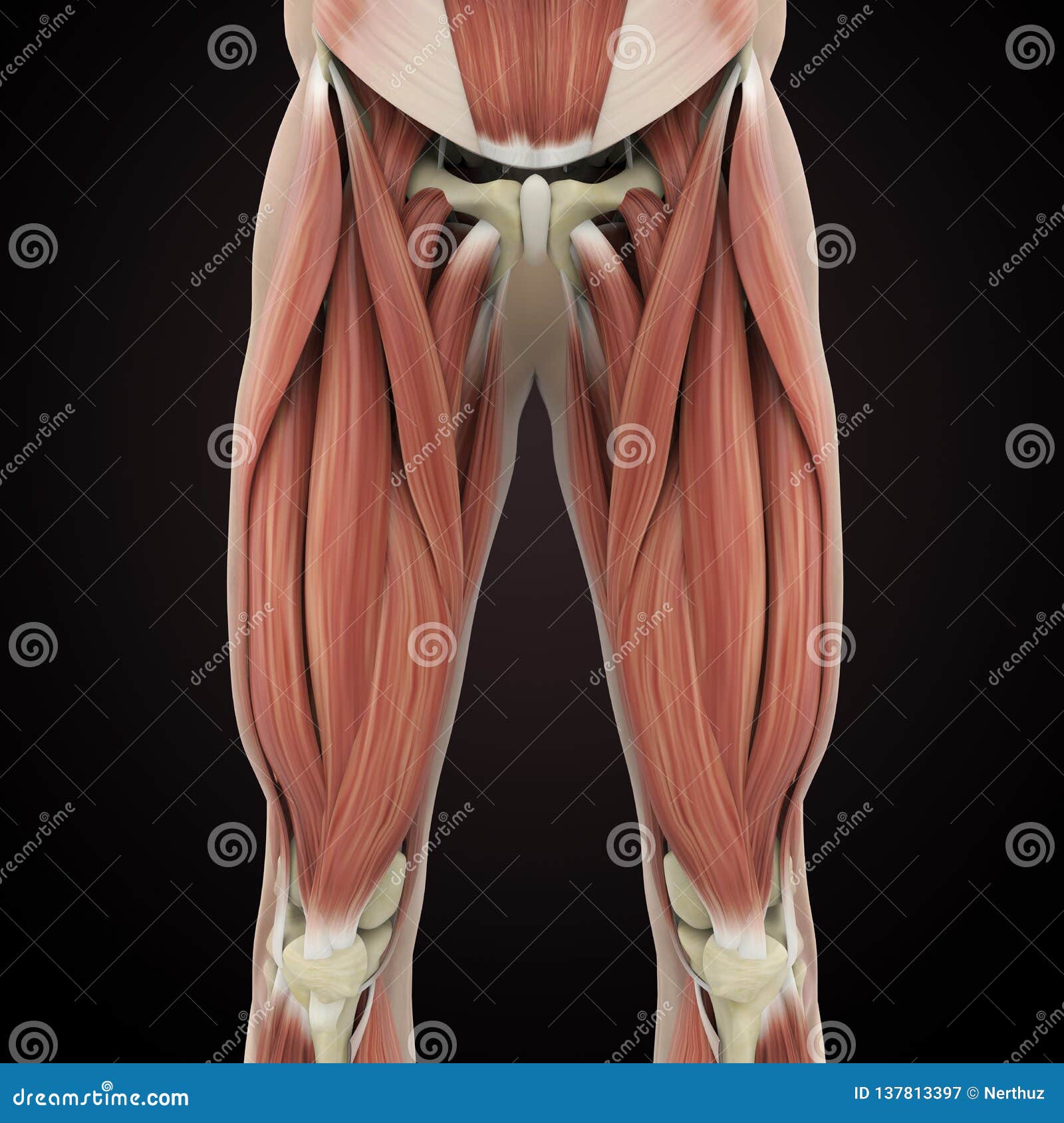
Upper Legs Muscles Anatomy Stock Illustration Illustration Of Iliacus 137813397 from thumbs.dreamstime.com Current techniques have tended to anatomical reconstruction of the lcl, pt and pf. When a muscle contracts, the tendon pulls on the bone causing the joint to move. Spicermanyt at checkout for 40% off this tutorial! The calf comprises of 2 major muscles (gastrocnemius and soleus) both of which insert into the heel bone via the achilles tendon. Palmar region , arteries (illustrations: Tendons are thick bands of tissue that connect muscles to bone. Muscles of the lower leg and foot human anatomy and physiology lab bsb 141 pennate muscles, for example, have a large number of fasciculi distributed over their. A collection of anatomy notes covering the key anatomy concepts that medical students need to learn.
We study anatomy at the practical anatomy class we study the human body.
Mnemonics that can be used to remember the anatomy of the ankle tendons from anterior to posterior as they pass posteriorly to the medial malleolus of the tibia under the flexor retinaculum in the tarsal tunnel include: An anatomical and biomechanical study. Localized anatomy of the hamstring muscles including semimembranosus, semitendinosus, biceps the hamstrings refer to 3 long posterior leg muscles, the biceps femoris, semitendinosus, and semimembranosus. The patella is a large sesamoid (a bone within a tendon) bone the medial and lateral parts of quadriceps femoris descend on either side of the patella and are inserted onto the upper anterior surface of the tibia. Muscle/tendon inflammation and pain along anterio… A collection of anatomy notes covering the key anatomy concepts that medical students need to learn. Spicermanyt at checkout for 40% off this tutorial! Tendons are cords made of tough tissue, and they work as special connector pieces between bone and muscle. Related online courses on physioplus. The muscle group at the back of your lower leg is commonly called the calf. Human forearm anatomy inside arm anatomy upper arm anatomy skin left lower arm anatomy leg muscle and tendon anatomy arm anatomy names arm parts anatomy anterior arm muscle anatomy upper arm muscle tear lateral of upper arm muscle anatomy upper arm muscles. We speak of the upper extremities (arms) and the lower extremities (legs). Collectively, the muscles in this area plantarflex and invert the foot.